Résumé

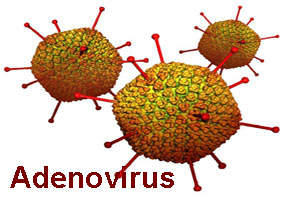
L'hépatite à corps d'inclusion (IBH) associée à l'infection par l'aviadénovirus aviaire (FAdV) a une distribution mondiale, en particulier chez les poulets de chair âgés de 3 à 5 semaines, entraînant des pertes économiques importantes de l'industrie avicole. Dans la présente étude, nous avons évalué la pathogénicité du virus FAdV- sérotype 11 isolé au Maroc (souche MOR111115) chez les embryons de poulets exempts d’agent pathogène (SPF). Le FAdV (titre 10? / ml) a été inoculé à des œufs embryonnés SPF à travers la membrane chorioallontoique (CAM). La mortalité, les lésions macroscopiques et microscopiques des embryons ont été évaluées et la présence du virus a été confirmé par réaction de polymérisation en chaine (PCR). La mortalité embryonnaire cumulative de 100% a été observée 7 jours après l'infection (dpi). Les embryons inoculés étaient hémorragiques et le foie était hypertrophié et friable, avec une décoloration jaune à verdâtre. Au microscope, nous avons mis en évidence une nécrose multifocale des groupes d'hépatocytes avec la présence de corps d'inclusion intra-nucléaires basophiles et éosinophiles dans les hépatocytes. La présence du virus a été confirmée par PCR conventionnelle basée sur le gène hexon à partir des prélèvements du foie, qui constitue l’organe cible des infections à FAdV. Il s'agit de la première étude de la pathogénicité de l'aviadénovirus aviaire dans les embryons de poulet SPF au Maroc.


Mots clés: Adénovirus aviaire, Hépatite à corps d’inclusion, Pathogénicité, Embryon de poulet

Introduction

Inclusion body hepatitis (IBH) associated with fowl aviadenovirus (FAdV) infection has a worldwide distribution (Schachner et al., 2018), especially for broiler chickens aged 3 to 5 weeks, causing significant economic losses to poultry industry. The IBH is caused by several serotypes of fowl adenovirus (McFerran et al., 2003). FAdVs are classified within the Aviadenovirus genus, family of Adenoviridae, and are further classified into five species (FadV-A to FAdV-E) and 12 serotypes (FAdV-1 to 8a and 8b to 11) based on cross neutralization assay (Benko et al., 2000; Hess, 2000).

In Morocco, outbreaks of IBH in chickens were reported for the first time in 2013 (Mouahid et al., 2013). Diagnosis was based on postmortem and histopathological examinations, which revealed enlarged and pale liver with the presence of basophilic intranuclear inclusion bodies in hepatocytes. Thereafter, several further cases of IBH in 2 to 3 week-old broiler chickens were detected in 2014 & 2015 based on macroscopic and microscopic changes (Said, 2015) followed by one outbreak that has been described in broiler breeders in 2017 (Salek et Elhoudfi, 2017). In 2018, the FAdV from broiler poultry was characterized and found belonging to FAdV-11 and FAdV-8a (Redondo et al., 2018). In 2019, FAdV was successfully isolated in CEF cell cultures and in specific pathogen free (SPF) chicken embryonated eggs from the liver samples collected from affected broiler and breeder broiler chickens. Those isolates were characterized and found belonging to FAdV-11 (Abghour et al., 2019).

The stand-alone pathogenicity of FAdVs had long been disputed, given the ubiquity of the viruses versus sporadic outbreaks. However, taking into account the increase of the cases in the field, numerous pathogenicity studies with FAdVs from outbreaks substantiated the primary etiologic role of particular strains, notably FAdV-4 the causative agent of hepatitis-hydropericardium syndrome and FAdV-2, -8a, -8b and -11 serotype (inclusion body hepatitis) (Schachner et al., 2018; Wei et al., 2019).

Chicken embryonated eggs (CEE) are proven approach for FAdV pathogenicity studies with several routes of inolculation through either chorioallantoic membrane (CAM), yolk sac or intravascular route (Cowen, 1988; Alemnesh et al., 2012; Maartens et al., 2014). However, the CAM route of inoculation of embryonated eggs was found more sensitive for adenovirus isolation (Kawamura et al., 1964). Furthermore, Some FAdV strains have been found to show different pathogenicity in chicken embryos (Lim et al., 2011; Almnesh et al., 2012; Joubert et al., 2014; Qing et al., 2017; Qinghua et al., 2018; Norfitriah et al., 2018). Therefore, the purpose of the present study was to investigate the pathogenicity of the recently isolated virulent FAdV-11 field strain (MOR 111115) on SPF Chicken embryonated eggs.

Materials and Methods

Chicken embryo fibroblast (CEF) cultures

CEF cultures were prepared from 9 to 11 day-old SPF chicken embryos according to standard procedures. Eagle’s growth medium (MEM) supplemented with 10% foetal calf serum, 100 U/ml penicillin and 0.1 mg/ml streptomycin were used. A monolayer of the CEFs was formed after about 24h of incubation at 37˚C under 5% CO2.

Avian adenovirus field strain

The FAdV isolate used in the present study (MOR 111115) was isolated from a liver sample during an outbreak of IBH on a commercial broiler chicken in Morocco in 2015 where clinical signs, necropsy and histological lesions characteristic of IBH infection were recorded. The genotyping revealed that the isolate belongs to serotype 11 (Abghour et al., 2019).

Preparation of virus inoculum

Liver samples from the infected chicken was collected and processed by three times frozen and thawed and then homogenized in a suspension of sterile phosphate buffered saline (PBS) 10% containing 200 IU/ml penicillin and 0.2 mg/ml streptomycin. The suspension was centrifuged at 2000g for 10 min at 4°C. The supernatant was filtered through 0.45 µm and 0.2 µm filter. 500 µl of homogenate was inoculated on confluent monolayer of the CEFs. The appearance of a cytopathic effect (CPE) characteristic of FAdV infection was monitored daily using an inverted microscope and Polymerase chain reaction (PCR) was performed to confirm the absence of contamination by NDV, IBV and H9-AIV. The third passage of the strain was kept at - 20°C for the next step of the study.

Determination of tissue culture infection doses (TCID50)

The TCID50 value of the virus strain were determined in 24-well plates covered with CEF cultures. The CEFs were infected with tenfold dilutions of virus from 10-1 to 10-7, three wells were made for each dilution and three wells for the negative control. The plates were incubated at 37°C with 5% CO2 and the cells were examined daily for the appearance of CPE. After seven days of incubation, the TCID50 was determined using the method of reed and Muench (1938).

Pathogenicity assessment of the FAdV isolate in SPF chicken embryos

Fifteen 10-days-old SPF embryos were inoculated with 100 µl of 105 TCID50/ml of CEF cell cultured virus via the chorioallantoic membrane (CAM) route. Five embryos were inoculated with sterile MEM as control. The eggs were incubated in humidified atmosphere (55%) at 37°C for 7 days. The embryos were candled daily to determine mortality. Embryos mortalities, which occurred before 24 h, were regarded as non-specific and discarded. All embryos, which died after 24 h post-inoculation, as well as those which survived until experiment termination, were harvested and necropsied for gross and histological examinations.

Gross and histological examination

On necropsy, the gross lesions were recorded and samples of liver were fixed in 10% neutral buffered formalin (NBF) for histological examination. The tissues were embedded in paraffin blocks according to standard methods, and cut into 4 μm sections, which were stained with hematoxylin and eosin (H and E) and examined under light microscope for lesions associated with FAdV infection.

DNA extraction

The embryos livers were processed for extraction of viral DNA using the Macherey Nagel Kit (Nucleospin Tissue, Germany) according to the manufacturer’s instructions but without preparing and pre-lyse sample steps.

Conventional PCR

Amplification of hexon gene was performed using published primers, HexF1, 5’GAYRGYHGGRTNBTGGAYATGGG-3’ and HexR1, 5’-TACTTATCNACRGCYTGRTTCCA-3’ (Mase et al., 2014). PCR reaction was performed as described previously (Abghour et al., 2019).

Results

Determination of the TCID50

After CPE was observed in infected CEF culture cells, the TCID50 of the MOR 111115 strain was found to be 105/ml using the reed –Muench method.

Mortality and gross lesions of SPF chicken embryos

The FAdV isolate MOR 111115 caused 100% cumulative embryonic mortality by day 7 post infection. The cumulative mortality of infected embryos revealed 13% embryos died at day 2 pi followed by 26% at 3 pi, 40% at 5-day pi, 66% at 6-day pi and 100% at day 7 pi (Figure 1).There was no mortality in control embryos throughout the experiment.

Upon necropsy, all uninfected embryos were normal without any changes in liver and CAM; however, at 2 to 7 day pi, the inoculated embryos were hemorrhagic, and showed enlarged livers, with either yellow to reddish foci or diffuse greenish discoloration (Figure 2A). At 6 and 7-day pi, the infected embryos were smaller than those of the control group (Figure 2B). The CAM was opaque, friable, hemorrhagic and thickened at 6 and 7-day pi (Figure 2C). The CAM of control embryos was thin with transparent membrane (Figure 2D).

Histopathological lesions

In control embryos, there were no changes in livers throughout the trial. Histological changes were observed in livers of inoculated embryos from 3 to 7 day pi. Multifocal necrosis of hepatocytes were detected with the presence of basophilic intra-nuclear inclusion bodies (Figures 3 and 4) some other inclusions were eosinophilic. The lesions were more extensive in liver at day 7 pi with congestion, hemorrhages and infiltration of inflammatory cells.

Virus detection by PCR

The PCR products visualized in agarose gel electrophoresis showed the presence of amplified DNA products of 800 bp in all DNA extracted from the liver samples of inoculated chicken embryos. This result confirms the presence of hexon protein gene specific for fowl adenovirus.

DISCUSSION

In recent years, FAdV-11 infection has been very common in commercial broiler and broiler breeder flocks in Morocco (Mouahid et al., 2013; Salek et El Houadfi, 2017; Redondo et al., 2018; Abghour et al., 2019). FAdV-11 induces IBH characterized by sudden onset of mortality ranging from 5% to 30%, which result in severe economic losses. IBHV has been isolated and characterized however, its pathogenicity was still unknown. Thus, the present work aimed to assess and determine for the first time the pathogenicity of the Moroccan isolate of FAdV-11(MOR 111115) in SPF chicken embryonated eggs based on mortality, gross and histological changes along with the confirmation of the presence of the virus by conventional PCR in liver of challenged embryos. It was demonstrated that the isolate is highly pathogenic in embryos with 100% cumulative mortality of embryos by day 7 pi. The highest cumulative mortality was recorded at 5 (40%), 6 (66%) and 7 (100%) day pi. The present finding is compatible with previous results of other experiments in which FAdV infection caused mortality in all infected embryos (Almnesh et al., 2012; Norfitriah et al., 2018). In other study, the mortality rates of embryos were 80-87% for FAdV-2 and 65-80% for FAdV-8b (Joubert et al., 2014). This variation of mortality may be related to the route of inoculation, the virulence of the virus inoculum (Cowen, 1988; Mendelson et al., 1995; Mazaheri et al., 1998).

Gross lesions observed in the infected embryos were confined mainly in the livers and CAM with moderate to severe necrosis, hemorrhages and thickening of the CAM. In addition, thickening of the CAM was found associated with opaque lesions at 6 and 7 day pi. Those finding are compatible with those previously described by other workers (Cowen, 1988; Mazaheri et al., 1998; Almnesh et al., 2012; joubert et al., 2014; Qing et al., 2017; Norfitriah et al., 2018).

Microscopic lesions observed in livers ranged from mild to severe and occurred at 3 to 7 dpi which is similar to changes, described in earlier trials (Almnesh et al., 2012; Norfitriah et al., 2018; Qinghua et al., 2018). Numerous large basophilic intra-nuclear inclusion bodies were easily identified in hepatocytes and some few others were eosinophilic which may be explained by extensive viral replication and liver damage (Itakura et al., 1977; Riddell, 1987; Weissenbock et Fuchs, 1995).

Viral DNA was detected in all liver samples collected from dead embryos. These results confirm the presence and replication of FAdV in livers of the infected embryos. Similar results were obtained by Qinghua et al., (2018).

In addition, a correlation between mortality, gross lesions, histological finding and viral DNA detection in liver of embryos infected with FAdV-11 was found at 6 and 7 dpi. The mortality of infected embryos was greatest at 6 and 7 dpi, gross lesions were recorded from 2 to 7 dpi, histological changes were recorded from 3 to 7 dpi and viral DNA was detected from 2 to 7 dpi.

CONCLUSION

This is the first Pathogenicity study demonstrating that FAdV-11(MOR 111115) isolated from outbreaks of IBH in Morocco, is highly pathogenic in SPF chicken embryos. This finding has confirmed also the possibility to use the liver of embryos for isolation and propagation of the virus.

REFERENCES

Abghour S., Zro K., Mouahid M., Tahiri F., TartaM., Berrada J., Kichou F. (2019). Isolation and characterization of fowl aviadenovirus serotype 11 from chicken with inclusion body hepatitis in Morocco. Plos One, 14: e0227004.

Almemnesc W., Hair-Bejo M., Aini I., Omar AR. (2012). Pathogenicity of fowl adenovirus in specific pathogen free chicken embryos. J. Comp. Pathol., 146: 223–229.

Benko M., Harrach B., Russel WC. (2000). Family Adenoviridae. In: van Regenmortel MHV et al, editors. VirusTaxonomy. Classification and nomenclature of viruses. Seventh Report of the International Committee on Taxonomy of Viruses: Academic Press, San Diego. 227–238.

Cowen B. S. (1988). Chicken embryo propagation of type I Avian Adenovirus, Avian diseases, 32: 347-352.

Hess M. (2000). Detection and differentiation of avian adenoviruses: A review. Avian Pathology, 29: 195–206.

Itakura C., Matsushita S., Goto M. (1977). Fine structure of inclusion bidies in hepatic cells of chickens naturally affected with inclusion body hepatitis. Avian Pathology, 6: 19-32.

Joubert H W., Aitchison H., Maartens L H., Venter E H. (2014). Molecular differentiation and pathogenicity of aviadenoviruses isolated during an outbreak of inclusion body hepatitis in south Africa. Journal of the South African Veterinary Association, 85:1.

Kawamura H., Shimizu F., Tsubahara H. (1964). Avian adenovirus: its properties and serological classification. National Institute of Animal Health Quarterly (Tokyo), 4:183-193.

Lim T H., Lee H J., Lee D H., Lee Y N., Park J K., Youn H N., Kim M S., Youn H S., Lee J B., Park S Y, Choi I S., Song C S. (2011). Identification and virulence characterization of fowl adenovirus in Korea. Avian Diseases, 55: 554-560.

Maartens L H., Joubert H W., Aitchison H., Venter E H. (2014). Inclusion body hepatitis associated with an outbreak of fowl adenovirus type 2 and Type 8b in broiler flocks in South Africa. Journal of the South African Veterinary Association, 85: 1-5.

Mase M., Mitake H., Inoue T., Imada T. (2009). Identification of group I-III avian adenovirus by PCR coupled with direct sequencing of hexon gene. J. Vet. Med. Sci., 71: 1239–1242.

Mazaheri A., Prusas C., Vob M., Hess M. (1998). Some strains of serotype 4 adenoviruses cause inclusion body hepatitis and hydropericardium syndrome in chickens. Avian Pathology, 27: 269-276.

McFerran J.B., Adair B.M. Group I adenovirus infection. In: Saif Y.M., Barnes H.J., Glisson J.R., Fadly A.M., McDougald LR et al. (2003). editors. Diseases of poultry, 11th Ed. Ames: Iowa State Press. pp. 214–227.

Mendelson C., Nothelfer HB. Monreal G. (1995). Identification and characterization of an avian adenovirus isolated from a ‘spiking mortality syndrome’ fied outbreak in broilers on the Delmarva Peninsula, USA. Avian Pathology, 24: 693-706.

Mouahid M., Bengoumi M., Kichou F. (2013). Case report of recent outbreak of inclusion body hepatitis among broilers in Morocco. Abstract. Proc. 18th World Veterinary Poultry Association Congress, Nantes, France. pp. 562.

Norfitriah M S., Hzir-Bejo M., Omar A R., Aini I., Nuruifiza M I. (2018). Malecular detection and pathogenicity of fow adenovirus isolated from disease outbreak in commercial layer farm. International Journal of Agricultural Sciences and Veterinary Medicine, 6: 73-84.

Oliver-Ferrando S., Dolz R., Calderón C., Valle R., Rivas R., Pérez M., Biarnés, M., Blanco A., Bertran K., Ramis A., Busquets N., Majó N. (2017). Epidemiological and pathological investigation of fowl aviadenovirus serotypes 8b and 11 isolated from chickens with inclusion body hepatitis in Spain (2011-2013). Avian Pathology, 46:157-165.

Qing P., Linlin L., Yongqiang W., Yanping Z., Xiaole Q., Changjum L., Yulong G., Xiaomei W., Hongyu C. (2017). The first whole genome sequence and pathogenicity characterization of fowl adenovirus 4 isolated from ducks associated with inclusion body hepatitis and hydropericardium syndrome. Avian Pathology, 46: 571-578.

Qinghua H., Xinxin M., Xiaoying H., Yanyan H., Shaohua Y., Lin Z., Ning C., Chuantian X. (2018). Pathogenicity and complete genome sequence of a fowl adenovirus serotype 8b isolate from China. Poultry Science, 98:573-580.

Redondo H., Sarabia FJ., Ait Tahala M., Bensassi Y., Gil I., Elbachir Erraji, et al. (2018). Characterization of strain of fowl adenoviruses circulating in Morocco. Poult. Sci., 97: 4057-4062.

Riddell C. (1987). Avian histopathology. American Association of Avian Pathologists, Kennett Square, Pennsylvania. pp. 7-17.

Said A. (2015). Étude clinico-pathologique de cas d’hépatite à inclusion chez la volaille entre 2012 et 2015. Thesis. Institut Agronomique et Vétérinaire Hassan II, Rabat, Morocco.

Salek M., El Houadfi M. (2017). The first case and description of FAdV-11 causing high mortalities in very young broiler breeders in Morocco. Abstract. Proc. 19th World Veterinary Poultry Association Congress, Edinburgh, UK.

Schachner A., Matos M., Grafl B., Hess M. (2018). Fowl adenovirus-induced diseases and strategies for their control – a review on the current global situation, Avian Pathology, 47: 111-126.

Wei Z., Liu H., Diao Y., Li X., Zhang S., Gao B., Tang Y., Hu J., Diao Y. (2019). Pathogenicity of Fowl Adenovirus (FAdV) Serotype 4 Strain SDJN in Taizhou Geese. Avian Pathology, 48:477-485.

Weissenbock H., Fuchs A. (1995). Histological and ultrastructural characterization of hepatic intranuclear inclusion bodies in psittacine birds and pigeons. Avian pathology, 24: 507-521.