Caractérisation moléculaire du virus des nervures jaunes nécrotiques de la betterave (BNYVV) infectant la betterave à sucre au Tadla
Résumé
La betterave sucrière est la principale source de sucre au Maroc. Les principales zones de production de la betterave à sucre sont Doukkala, Tadla, Gharb, et Moulouya. La rhizomanie, causée par le virus des nervures jaunes nécrotiques de la betterave (BNYVV, genre Benyvirus), a été décrite pour la première fois en Italie et s'est propagée dans la plupart des régions productrices de la betterave sucrière. Dans la région de Tadla, la rhizomanie est l'un des principaux problèmes phytosanitaires majeurs pour la culture de la betterave sucrière. En 2010, cinq isolats de différentes localités de la région de Tadla ont été collectés pour des études de caractérisation moléculaire par le séquençage du gène de protéine de la capside (CP). Les résultats obtenus ont montré que les isolats marocains étaient dans le groupe B de BNYVV; ces isolats ont montré une grande similarité nucléotidique avec l'isolat Belge B2 (AY696077), avec une homologie de séquence de 100 %. Au mieux de nos connaissances, il s'agit de la première caractérisation moléculaire des isolats de BNYVV au Maroc.
Mots-clés: Rhizomanie, betterave sucrière, caractérisation moléculaire, gène de la protéine de la capside, Maroc.
Téléchargements
INTRODUCTION
Production of sugar beet is the most important sugar crop in Morocco. With an area of cultivation of 58.400 ha, it produces 3.6 million tons of roots (Anonymous, 1015). The main sugar beet production areas in Morocco are Doukkala, Tadla, Gharb, and Moulouya. Sugar beet production is suffering from many problems, especially from diseases and pests. Very scarce information is available on viral diseases of sugar beet, especially on Beet necrotic yellow vein virus (BNYVV, genus Benyvirus), the causal agent of Rhizomania, which is one of the major phytosanitary problems for the sugar-beet industry worldwide. Heavy infection with rhizomania can cause a yield loss of up to 70% (Johansson, 1985) and a decrease in sugar content from 16-18% to less than 7% (Bongiovanni and Lanzoni, 1964).
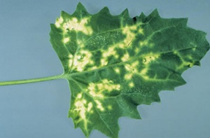
BNYVV is transmitted by soil-borne plasmodiophorid (protozoan) vector, Polymyxa betae Keskin (Abe and Tamada, 1986; Tamada, 2002). The virus is cystosorus-borne and survives many years in soils even without susceptible plants (Tamada, 2002). The disease appears as foci (patches) in low laying areas of sugar beet fields. The infected sugar beet plant shows mild to severe chlorosis particularly in vascular bundle, followed by venial necrosis (necrotic yellow vein). The virus also produces characteristic symptoms on roots by exhibiting proliferation of lateral rootlets (rhizomania) and stunting (Tamada, 2002).
BNYVV has a multipartite single-stranded RNA genome with all natural isolates containing four RNA species, although some isolates have an additional RNA-5 (Tamada et al., 1989). Three major serologically indistinguishable BNYVV groups named type A, B and P have been identified (Kruse et al., 1994; Koenig et al., 1995; Miyanishi et al., 1999; Koenig and Lennefors, 2000). BNYVV A type isolates are distributed worldwide, whereas the B type isolate is prevalent in limited areas of Europe, mainly in France and Germany, but occurs also in China and Japan (Miyanishi et al., 1999; Sohi and Maleki, 2004; Schirmer et al., 2005; Li et al., 2008). BNYVV A and B types typically contain only RNAs 1 to 4. BNYVV P type isolates that contain RNA-5 are detected only in France, Kazakhstan and in the UK (Koenig et al., 1997; Koenig and Lennefors, 2000; Harju et al., 2002; Ward et al., 2007). P type isolates appear to be more virulent than both the A and B types (Heijbroek et al., 1999) and may evade activation of plant defense responses.
BNYVV was first reported from Italy (Canova, 1959) and Tamada and Baba (1973) identified the causal agent of this disease as a virus and later they called it BNYVV from the leaf symptoms it induces. It has since been reported from many sugar beet producing countries all over the world (Chiba et al., 2011). In Morocco, Rhizomania was first detected in 2004 in Tadla region, and then the presence of the virus was confirmed in Doukkala region (EPPO, 2005).
Although BNYVV has occurred in Morocco since 2004, no attempt has been carried out to characterize the local virus type. Therefore, the present study aims to characterize local BNYVV isolates.
MATERIAL AND METHODS
Five BNYVV sources were collected from different locations in the Tadla region (Afourer, Beni Amir, Dar Ouled Zidouh, Ouled M’Bark and Souk Sebt) and were used to perform RNA extractions using RNaesy Plant Mini Kit (Qiagen, Germany) according to the manufacturer’s instructions; then, tested by RT-PCR using specific primers to amplify a 567-bp fragment of the coat protein (CP) gene of BNYVV (Schirmer et al., 2005). The PCR program used was 94°C for 3 min, 35 cycles of 94°C for 1 min, 61°C for 1 min and 74°C for 1 min, followed by final extension at 72°C for 10 min. PCR products were electrophoresed in 1.2% agarose gel in 1X TAE buffer and stained with ethidium bromide (25 ng/ml) and visualized on UV trans-illuminator. Fragment sizes were determined by comparison with a 1 Kb DNA standard marker (GeneRulerTM DNA Ladder, 0.5 ug/ul).
PCR products corresponding to the CP (567 bp) gene, from five local BNYVV isolates form Tadla region (TDL1, TDL2, TDL4 and TDL5), were purified using a DNA extraction kit (Fermentas, Lithuania) and sequenced. Sequencing was carried out in two directions and each sample was sequenced twice. The obtained sequences were aligned using the BioEdit software version 7.0.9 (Hall, 1999) and the MEGA software version 5.05 (Tamura et al., 2011). The CP gene sequence of one Moroccan isolate from Tadla region (RHZ-TDL-Mor) was deposited in the GenBank database under the accession number KY771166. RHZ-TDL-Mor was then confronted with other strain references available in the NCBI database. Calculation of pair wise nucleotide distances between sequences and clustering were performed using the phylogenetic and molecular evolutionary genetic analysis software MEGA version 5.05 for the estimation of the nucleotide homology (Tamura et al., 2011).
RESULTS AND DISCUSSION
Multiple alignments and the sequence identity matrix of the CP gene showed a high sequence similarity among the five sequenced Moroccan BNYVV isolates in the Tadla region (100 % nucleotide identity). Thus, a low genetic diversity exists within the Moroccan BNYVV population; suggesting that the virus was not introduced into Morocco from multiple sources. The obtained results showed that the Moroccan isolate RHZ-TDL-Mor (KY771166) was in the B type of BNYVV and showed the highest nucleotide similarity with the Belgian isolate B2 isolate (AY696077), with a sequence homology of 100% (Figure 1).
RHZ-TDL-Mor clustered with Belgian (AY696077) and Deutsch (AB56306) isolates indicating a possible common entrance or introduction from these regions. We also found that coat protein gene of local RHZ-TDL-Mor isolate was closely related to other sequences of different BNYVV isolates available in the database with a sequence homology ranging from 96.2% to 98.9%; supporting the idea that coat protein is a relatively stable sequence in BNYVV genome.
Comparisons based on CP-encoding nucleotide sequences indicated that percentage identity is highly conserved for all the isolates reported worldwide, suggesting that either the virus has a very stable genome or this might be the incidence of a recent introduction of the virus in different sugar beet growing areas. The result is quite in agreement with the findings of Bouzoubaa et al., (1987) where high level of genome conservation was reported for BNYVV reported isolates.
Rhizomania is one of the most economically important diseases affecting sugar beet, and is widely distributed in most sugar beet growing areas of the world. The studies on BNYVV worldwide revealed that there are three pathotypes; A-type, B-type, and P-type (Kruse et al., 1994; Koenig et al., 1995; Schirmer et al., 2005). The A-type is distributed throughout most sugar beet growing regions of the world and is, so far, the only form present in the Tadla region.
Fields remain infested with BNYVV indefinitely by Polymyxa betae cystosori that remain dormant for up to 25 years. It has been reported that the environmental conditions and cultural practices do not alter the viability of the virus inside the resting spores (Tamada, 2002; Rush, 2003). Therefore, rotation to non-host crops or lengthening rotations is ineffective at reducing disease incidence, and the only viable means of control has been natural host-plant resistance. Indeed, the virus detection and timely diagnosis is of utmost importance.
CONCLUSION
BNYVV is one of the most economically important viruses infecting sugar beet in Morocco. Molecular characterization studies showed low genetic diversity among BNYVV isolates in the Tadla region. Also, these local isolates were all of the BNYVV B type and showed the highest nucleotide similarity with the Belgian BE isolate. Results of the present study are important in terms of future efforts aiming at restraining the spread of BNYVV to new areas. As with other soil-borne viruses, efforts to eradicate BNYVV from newly contaminated fields could be very difficult if not impossible without abandoning cultivation of susceptible crops for years. In areas where BNYVV occurs, introduction of rhizomania-resistant sugar beet cultivars is the most important option whose sustainability depends on the genetic variability of the local BNYVV populations. This study provides information for planning future strategies to control rhizomania in Morocco.
Aspects which require further investigations include determining the density of Polymyxa betae carry BNYVV, and the stimulation of cystosori in the soil to release zoospores in absence of the host or the needed trap plants to attract zoospores before planting sugar beet in infected soils. Additionally, more investigations are needed to determine the predominant BNYVV type in the other beet sugar cultivation areas. Answers to these questions may help to develop control strategies against BNYVV and other viruses transmitted by plasmodiophorid vectors.
REFERENCES
Abe H., Tamada T. (1986). Association of Beet necrotic yellow vein virus with isolates of Polymyxa betae Keskin. Annals of the Phytopathological Society of Japan 52: 235-247.
Anonymous. (2015). Ministère de l’Agriculture et de la Pêche Maritime. http://www.agriculture.gov.ma/.
Bongiovanni G.C., Lanzoni L. (1964). La rizomania della bietola. Progresso Agricolo 2: 209-220.
Bouzoubaa S., Quillet L., Guilley H., Jonard G., Richards K. (1987). Nucleotide sequence of Beet necrotic yellow vein virus RNA-1. Journal of General Virology 68: 615-626.
Canova A. (1959). Appunti di patologia della barbabietola. Informatore Fitopatologico 9: 390-396.
Chiba S., Kondo H., Miyanishi M., Andika I.B., Han C., Tamada T. (2011). The evolutionary history of Beet Necrotic yellow vein virus deduced from genetic variation, geographical origin and spread, and the breaking of host resistance. Molecular Plant - Microbe Interaction 24: 207-218.
EPPO (2005). Premier Signalement du Beet necrotic yellow vein benyvirus au Maroc. Service d’Information, No. 7.
Johansson E. (1985). Rhizomania in sugar beet-a threat to beet growing that can be overcome by plant breeding. Sveriges Utsӓdesfӧrenings Tidskrift 95: 115-121.
Hall T.A. (1999). BioEdit: a user-friendly biological sequence alignment editor and analysis program for Windows 95/98/NT. Nucleic Acids Symposium Series 41: 95-98.
Harju V.A., Mumford R.A., Bockley A., Boonham N., Clover G.R.G., Weekes R., Henry C.M. (2002). Occurrence in the United Kingdom of Beet necrotic yellow vein virus isolates which contain RNA 5. Plant Pathology 51: 811.
Heijbroek W., Musters P.M.S., Schoone A.H.L. (1999). Variation in pathogenicity and multiplication of Beet necrotic yellow vein virus (BNYVV) in relation to the resistance of sugar-beet cultivars. European Journal of Plant Pathology 105: 397-405.
Koenig R., Luddecke P., Kaeberle AM. (1995). Detection of Beet necrotic yellow vein virus strains, variant and mixed infections by examining single-strand confirmation polymorphisms of immunocapture RT-PCR products. Journal of General Virology 76:2051-2055.
Koenig R., Lennefors B.L. (2000). Molecular analyses of European A, B and P type sources of Beet necrotic yellow vein virus and detection of the rare P type in Kazakhstan. Archives of Virology 145: 1561-1570.
Koenig R., Haeberle A., Commandeur U. (1997). Detection and characterization of a distinct type of Beet necrotic yellow vein virus RNA 5 in sugar beet growing areas in Europe. Archives of Virology 142: 1499-1504.
Kruse M., Koenig R., Hoffmann A., Kaufmann A., Commandeur U., Solovyev A.G., Savenkov I., Burgermeister W. (1994). Restriction fragment length polymorphism analysis of reverse transcription-PCR products reveals the existence of two major strain groups of Beet necrotic yellow vein virus. Journal of General Virology 75:1835-1842.
Li M., Liu T., Wang B., Han C., Li D., Yu J. (2008). Phylogenetic analysis of Beet necrotic yellow vein virus isolates from China. Virus Genes 36: 429-432.
Miyanishi M., Kusume T., Saito M., Tamada T. (1999). Evidence for three groups of sequence variants of Beet necrotic yellow vein virus RNA 5. Archives of Virology 144: 879-892.
Rush C.M. 2003. Ecology and epidemiology of benyviruses and plasmodiophorid vectors. Annual Review of Phytopathology 41: 567-592.
Schirmer A., Link D., Cognat V., Moury B., Beuve M., Meunier A., Bragard C., Gilmer D., Lemaire O. (2005). Phylogenic analysis of isolates of Beet necrotic yellow vein virus collected from worldwide. Journal of General Virology 86: 2897-2911.
Sohi H.H., Maleki M. (2004). Evidence for presence of types A and B of Beet necrotic yellow vein virus (BNYVV) in Iran. Virus Genes 29: 353-358.
Tamada T. (2002). Beet necrotic yellow vein virus. CMI/AAB Descriptions of Plant Viruses N° 391. Association of Applied Biologists, Wellsbourne, United Kingdom.
Tamada T., Baba T. (1973). Beet necrotic yellow vein virus from rhizomania-affected sugar beet in Japan. Annals of the Phytopathological Society of Japan 39: 325-332.
Tamada T., Shirako Y., Abe H., Saito M., Kigushi T. and Harada T. (1989). Production and pathogenicity of isolates of Beet necrotic yellow vein virus with different number of RNA components. Journal of General Virology 70: 3399-3409.
Tamura K, Peterson D, Peterson N, Stecher G, Nei M., Kumar S. (2011). MEGA5: Molecular Evolutionary Genetics Analysis using Maximum Likelihood, Evolutionary Distance, and Maximum Parsimony Methods. Molecular Biology and Evolution. http://www.kumarlab.net/publications.
Ward L., Koenig R., Budge G., Garrido C., McGrath C., Stubbley H., Boonham N. (2007). Occurrence of two different types of RNA-5-containing Beet necrotic yellow vein virus in the UK. Archives of Virology 152: 59-73.
Publié-e
Numéro
Rubrique
Licence

Revue Marocaine des Sciences Agronomiques et Vétérinaires est mis à disposition selon les termes de la licence Creative Commons Attribution - Pas d’Utilisation Commerciale - Partage dans les Mêmes Conditions 4.0 International.
Fondé(e) sur une œuvre à www.agrimaroc.org.
Les autorisations au-delà du champ de cette licence peuvent être obtenues à www.agrimaroc.org.